
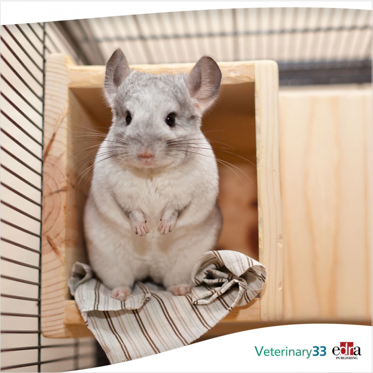
Praeputial calculi leading to pollakiuria in a chinchilla (Chinchilla lanigera)

A 3-year-old male intact 530g chinchilla was referred to the clinic due to a severely swollen genital region that had been developing progressively for 8months. Within one week of presentation, the animal showed pollakiuria and the urine ran along the coat instead of being passed in a stream.
On clinical examination of the genital region, solid, crepitant, non-sliding calculi were palpable over the entire length of the preputium up to the pelvic inlet. Directly caudal of the pelvic inlet was a displaceable, round structure. A radiographic examination revealed multiple round to cylindrical, mineral dense concrements in the caudodistal genital area. In addition, multiple tubular structures with heterogeneous contents were present, which were interpreted as intestinal loops because of a hernia inguinalis. The urinary bladder was moderately filled and free of calculi, as was the urethra. Under general anaesthesia with isoflurane and oxygen supplementation, extensive preputial lavage was performed. Brown, rough calculi were completely removed. Praeputial mucosa showed multifocal green debris and ulcerations, and low-grade haemorrhage was present at the tip of the penis. Both testicles as well as other possible prolapsed organs were freely reducible into the abdominal cavity, so the hernia was presumed clinically insignificant. The therapy was continued with enrofloxacin and buprenorphine, meloxicam and metamizole were started at recommended doses. The chinchilla recovered well after anaesthesia and showed normal urination with passing urine in a stream and resolution of pol- lakiuria. The owner was instructed to perform praeputial rinsing with 0.9% NaCl twice a day with subsequent installation of dexpanthenol containing eye ointment into the prepuce for 3days. Urolith analysis revealed 100% calcium carbonate.
In conclusion as a general recommendation, an increased water intake and avoiding the feeding of herbs, pellet food and vegetables high in calcium was advised.
 Comments
Related Articles

Copyright © 2026 - All Rights Reserved
ISSN 2768-198X
List
Add
Please enter a comment