
Diagnostic imaging, why compare the methods?
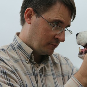
An interview with Professor Massimo Vignoli, University of Torino

Go beyond watertight compartments: an effective diagnostic approach cannot do without comparing methods, highlighting their pros and cons in clinical situations—an interview with the author of the Atlas of diagnostic imaging of dogs and cats.
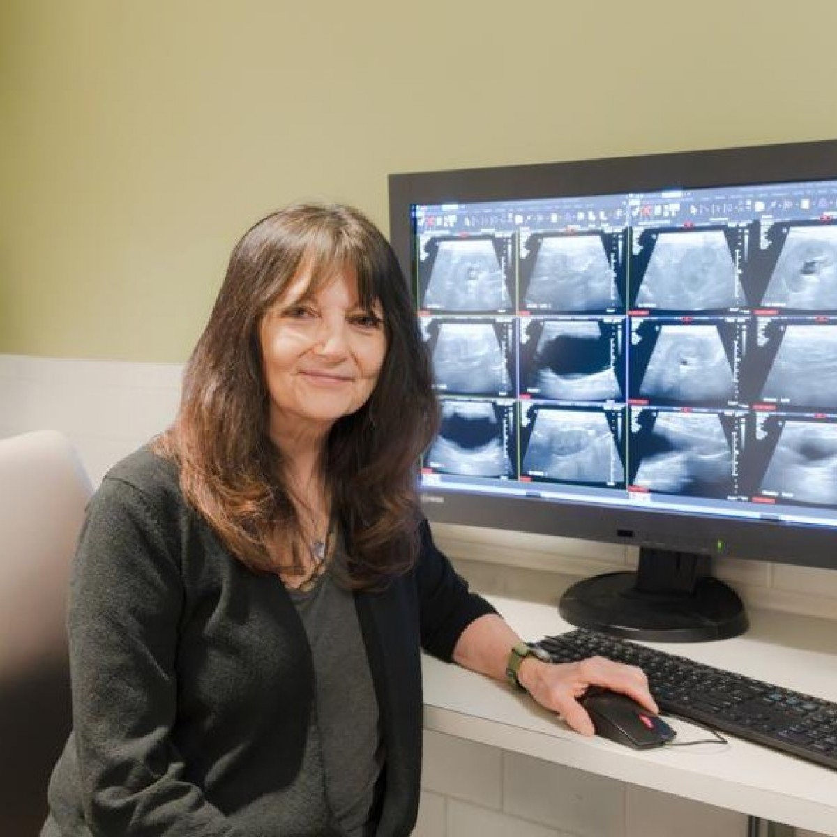
There is no map in this Atlas. But x-rays, ultrasounds, and imaging resonances, yes. The world that is being illustrated, image by image, is not a geographical space but a complex and fascinating universe that tells the story of feline and canine anatomy and the pathologies connected to them. And here, in an exploratory journey into the perfect and accomplished microcosm of every single organ and system, the “Atlas of Diagnostic Imaging of Dogs and Cats” leads the reader into the worlds in which to look. A heavy volume enriched with about 1,550 images, the process of writing it lasted more than three years and involved, together with the two editors, over 20 co-authors selected from international specialists in the field. In this interview, Professor Massimo Vignoli, author of the work with Dr. John Graham, tells us about the atlas.
"The atlas is the culmination of a project that brings together all the diagnostic imaging methods in a single volume, to compare them and illustrate from time to time, based on the clinical picture, which is more useful to adopt. In the past, I have organized courses with a synoptic approach. Bringing everything together in one book is very satisfying for me."
Q: Why create this book on diagnostic imaging now?
Vignoli: In the last 10 years, this has become one of the most important areas in both human medicine and veterinary medicine, as it recognizes collateral investigations that are fundamental in daily clinical practice and different for each clinical case. It is, therefore, a priority to deepen the topic, starting with comparing the various methods and highlighting their pros and cons.
Until now, literature has been limited to monographs on a single technique or, at most, two. In this book, we wanted to focus on the most common diseases (i.e., 70 to 80% of those treated daily in clinics) and then explain which imaging technique best suits the specific case. Sometimes an x-ray study is sufficient, while an ultrasound, a CT scan, or an MRI is more useful in other situations.
In this sector, it is essential to have a diagnostic approach methodology. For this reason, we used super specialists, all graduates of European or American veterinary colleges.
Q: What if a unique aspect of the atlas?
Vignoli: I would undoubtedly say the impressive iconographic apparatus and the choice to accompany it only with brief descriptive texts of the organs' diseases and the various systems, so that the images can speak for themselves.
Q: What is the audience you’re hoping to reach?
Vignoli: It was created to meet the needs of veterinary professionals conducting clinical activity. And, given the reported cases, it can also be a useful tool for students and specialist radiologists.
Q: Speaking of students, how much space is given to this specialty in universities?
Vignoli: Very little, but it’s due to a matter of time. Veterinary students typically learn the basics of radiology, ultrasound or tomography. More in-depth knowledge is gained at the post-degree level, where you can opt for a master's degree or pursue doctoral research doctorate at European or American colleges. And in this case, the path is tough. You must believe in it so much because it's four years of study and full-time work. But in the end, it will be worth it.
Q: Let's return to your commitment as editor; what are you working on next?
Vignoli: I am editing the Italian version of the atlas, which is scheduled to be published in fall 2023. This time, I no longer have the collaboration of a co-author, who, in the English version - in addition to the scientific contents - had taken care of the shape. At the same time, I dedicated myself to redefining the images. Now it will be up to me to bear the burden and the honor of carrying out the linguistic checks of the case.
About Professor Massimo Vignoli
Vignoli has a doctoral degree in veterinary sciences from the University of Ghent and he is a specialist in veterinary radiology. He graduated from the European College of Veterinary Diagnostic Imaging (ECVDI). Vignoli is a professor at the University of Torino, where he is also director of the second-level university master's degree program.
Learn more about the atlas: https://www.edrapublishing.com/home/600/atlas-of-diagnostic-imaging-of-dog-and-cat.html
 Comments
Related Articles

More news


Copyright © 2026 - All Rights Reserved
ISSN 2768-198X
List
Add
Please enter a comment