
Canine leishmaniasis in the U.S.

Leishmaniasis is one of the most common zoonoses worldwide and a serious public health concern, yet it is often overlooked and neglected in dogs in the U.S. With the number of cases rising in this country, veterinarians must become familiar with the clinical signs as well as proper diagnosis, treatment, and management.
In the past decade, several vaccines have been developed and used to prevent or aid in treatment of disease in Leishmania-endemic areas. Developing a program for screening dogs imported into the U.S. from Leishmania-endemic regions and ensuring that U.S. veterinarians have the information necessary to screen, identify, and treat patients with leishmaniasis is critical for preventing the spread of Leishmania.
According to the authors and the study published recently in Today's Veterinary Practice, Leishmaniasis has not been considered a significant concern to veterinarians in the U.S. because the sand flies capable of transmitting the parasite have been documented in only a few isolated parts of the country and prevalence of leishmaniasis in dogs native to the U.S. has been relatively low.
However, the number of cases of canine leishmaniasis has increased, from importation of dogs from Leishmania-endemic areas and local acquisition. Autochthonous cases have been reported in California, Georgia, Maryland, New York, North Dakota, Oklahoma and Texas.
What's contributing to the rise of canine leishmaniasis in the U.S.? The authors said vertical transmission, international travel and animal transportation, and climate change.
Like people, dogs, cats and other animals can be infected by a variety of species of Leishmania. The main reservoir hosts, however, are dogs, susceptible to different forms of disease, ranging from self-limiting cutaneous lesions to severe visceral disease that can be fatal if untreated.
Veterinarians should consider the disease as a potential diagnosis for dogs in North America, especially ill dogs that have been to or bred to any dog from a Leishmania-endemic region.
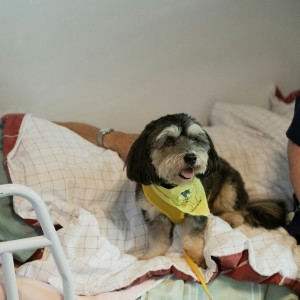
This article describes a case of canine leishmaniasis in a five-year-old spayed female Australian shepherd recently imported to the U.S. from a shelter in the Henan province of China.
According to the case report included in the study, on initial examination in China, the patient appeared healthy, except for being underweight and having moderate to severe dental tartar. Basic veterinary care was provided, including core vaccinations and occasional doses of flea protection. She progressively lost weight, and lameness developed and worsened over the nine months preceding her transport to the U.S.
One month before transport, progressively worsening lameness was noted. Radiographs showed marked soft tissue swelling around the right elbow with normal appearing carpi and long bones. No cause was identified. Immediately before transport, non–weight-bearing lameness developed on the left forelimb. Radiographs showed increased soft tissue swelling of the right elbow and mild right carpal soft tissue swelling. A chondroitin supplement was started. Because of the dog’s clinical decline, she was taken to a veterinarian immediately after arrival in the U.S.
A definitive diagnosis
A diagnosis of leishmaniasis was based on history, clinical signs and clinicopathologic abnormalities consistent with leishmaniasis, as well as the high titer and, ultimately, response to treatment. Although not necessary for a definitive diagnosis, and not done in this case study, identifying the organism in tissue supports the diagnosis.
Treatment
Considering the severity of the joint effusions and inflammatory component, treatment was started immediately pending diagnostic results. Treatment included gabapentin (20 mg/kg PO q8h) for discomfort, doxycycline (10 mg/kg PO q24h) to cover possible tick-borne infection and for its anti-inflammatory properties, and intra-articular injections of triamcinolone (3 mg) into both tarsi. Because of the marked proteinuria, the patient was given oral clopidogrel (1.5 mg/kg PO q24h) as an antithrombotic and oral telmisartan (1 mg/kg PO q24h) to lessen renal protein losses.
In response to the joint injections and supportive care, the patient’s lameness, fever, and azotemia resolved within 48 hours. After receiving the positive immunofluorescence antibody titer confirming a diagnosis of leishmaniasis, allopurinol (10 mg/kg PO q12h indefinitely) and meglumine antimoniate (100 mg/kg SC, divided into 2 equal doses q12h for 6 weeks) were prescribed.9
Outcome and future treatment
All clinical signs resolved within two to four weeks of starting allopurinol and meglumine antimoniate treatment, and after eight months, the proteinuria resolved. However, the Leishmania titer remain elevated.
Treatment with allopurinol will continue until titers significantly decline or become negative. If clinical signs return after the initial course of treatment, allopurinol therapy alone or with a course of miltefosine will be restarted. Blood work, urine, and Leishmania immunofluorescence antibody titer will be monitored annually for the rest of the patient’s life. If no clinical signs recur, she will enjoy her life free of medication.
Read more about additional diagnostic tests and other details: "Canine Leishmaniasis in the United States."
Diane Levitan and Jacqueline Finnegan. "Canine Leishmaniasis in the United States." Today's Veterinary Practice. 13 February 2023. Vol. 13, Number 2.
 Comments
Related Articles


More news


Copyright © 2026 - All Rights Reserved
ISSN 2768-198X
List
Add
Please enter a comment