
Pathologic and Immunohistochemical Findings in a Feline Aortic Body Tumor

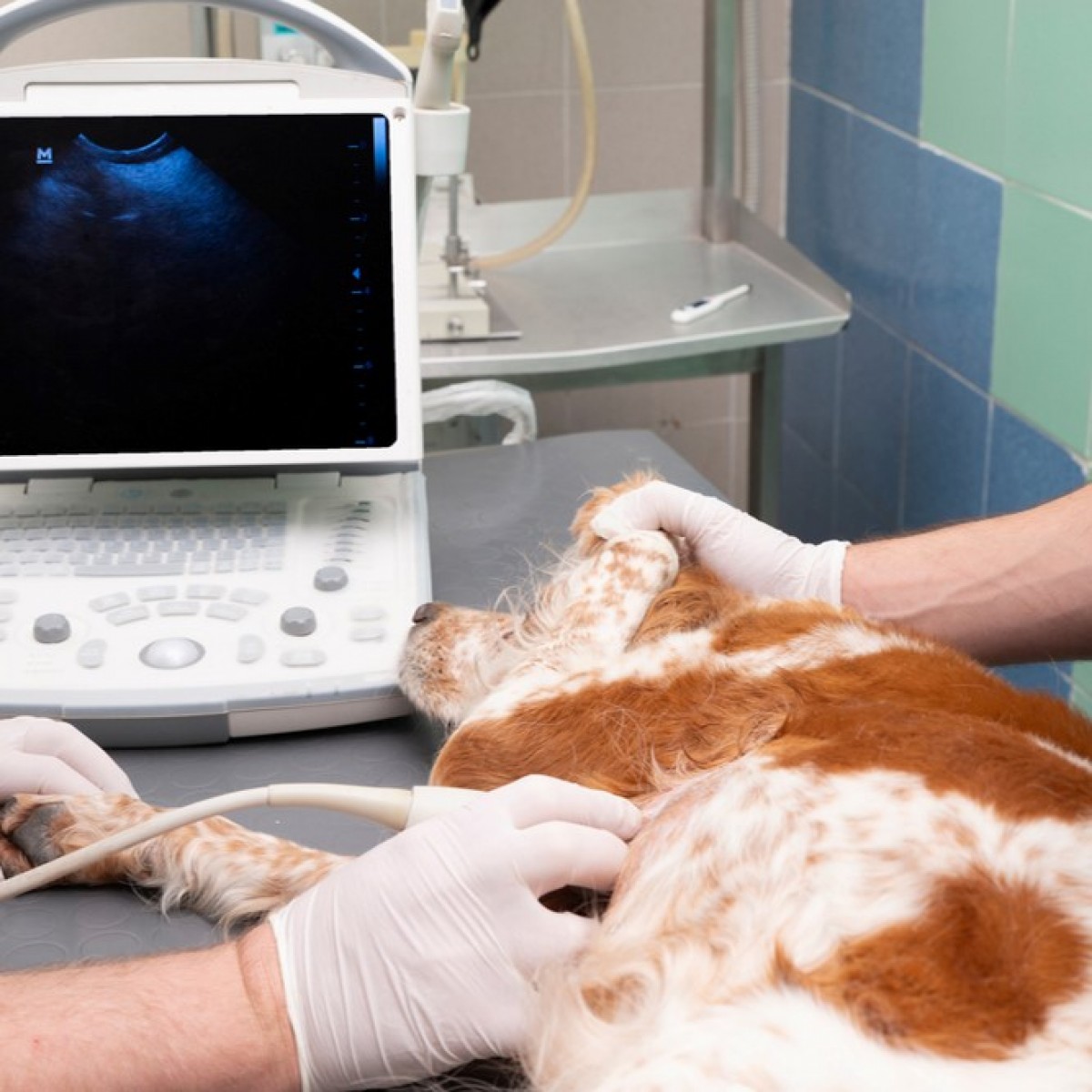
The presence of a heart-base tumor was diagnosed by ultrasound imaging in a 10-year-old, female, domestic shorthaired cat presenting with dyspnea and pleural effusion because of the presence of a modified transudate. Hematology and clinical chemistry were unremarkable. The owner elected euthanasia. At necropsy, a locally extensive, firm, multilobulated nodule surrounded the pulmonary vein. The tumor was composed of lobules of large polygonal cells separated by a fine fibrovascular stroma. Tumor cells infiltrated the myocardium, and neoplastic emboli were present, but no metastases were macroscopically detectable. Tumor cells were immunohistochemically positive for chromogranin A, for synaptophysin and, faintly, for neuron-specific enolase and negative for vimentin, cytokeratin, a smooth muscle actin, glial fibrillary acidic protein, thyreoglobulin, and calcitonin. Based on histologic and immunohistochemical findings, the diagnosis of chemodectoma was made.
Chemodectoma, or extra-adrenal nonchromaffin paraganglioma, is a neoplasm arising from chemoreceptor cells involved in the response to changes in blood pH, oxygen tension, and carbon dioxide content.4,13,20 Chronic hypoxia is probably involved in the pathogenesis of chemodectoma: a high prevalence of this tumor has been recorded both in humans and in cattle living in mountain areas1,21 and in brachicephalic dogs, which have an upper respiratory tract anatomic conformation that favors a chronic hypoxic status.13 Chemodectomas most commonly arise within the aortic or the carotid body 4,13,20 and, less frequently, in the glomus pulmonale,20 in the glandula suprarenalis,9 or in ectopic sites.2,7,8,15,20 Chemodectomas are particularly rare in cats: to date, two carotid body tumors,5,24 one cauda equina paraganglioma,8 and eight aortic body tumors3,6,11,12,19,22–24 have been reported in cats. Aortic body tumors are locally extensive, multilobulated masses, located within the pericardium at the heart base.4 They are usually encapsulated and organized in small lobules surrounded by a prominent fibrovascular stroma.4 Neoplastic cells commonly stain for neuron-specific enolase (NSE), chromogranin A (CgA), and synaptophysin (SY).7,8,15,18
Immunohistochemical characteristics of feline aortic body tumors, however, have not been described. We report the clinicopathologic and immunohistochemical features of a feline aortic body tumor.
A 10-year-old, female, domestic shorthaired cat was presented to the referring veterinarian due to severe inspiratory dyspnea and cyanosis. Clinical and radiographic examination revealed a severe thoracic effusion. Serology for feline immunodeficiency virus, feline leukemia virus, and feline coronavirus was negative. Hematology and clinical chemistry were unremarkable. Cytology of the pleural fluid was characterized by neutrophils, mostly nondegenerate, macrophages, and reactive mesothelial cells (modified transudate). After thoracocenthesis (150 ml of yellowish effusion was removed), radiography and ultrasound imaging of the chest revealed the presence of a 25-mm nodule, located at the heart base and surrounding the large vessels. Because of the poor prognosis, the cat was euthanatized. A full necropsy was performed by the referring veterinarian. Samples of tumor, large vessels, and myocardium were collected, fixed in 10% buffered formalin, routinely processed, and embedded in paraffin.
Read the full article here.
Authors: S. Paltrinieri, P. Riccaboni, M. Rondena, C. Giudice
Source: https://journals.sagepub.com/
 Comments
Related Articles


Copyright © 2026 - All Rights Reserved
ISSN 2768-198X
List
Add
Please enter a comment