
Tips for Mastering Veterinary Dental Radiology
Apart from having personnel trained in this matter on the team, it is advisable to follow these practical tips to obtain the best results.
Obtaining full mouth dental X-rays can seem like a very complicated process, especially in non-specialist centers where it is not done routinely. Apart from having personnel trained in this matter on the team, it is advisable to follow these practical tips to obtain the best results.
1. Synchronization
Full mouth radiographs should be obtained early in the procedure, immediately after the patient is anesthetized and stabilized. Doing it this way helps start the diagnostic process with some objective data. Once the radiographs have been examined, an abnormality will likely be found, and the client will need to be called for treatment approval. If waiting until everything else is done to obtain the X-rays, and the client does not respond when asked for treatment authorization, the patient's time will remain anesthetized may be unnecessarily extended.
2. Patient positioning
Patients should be positioned the same way each time an X-ray is obtained. With the sternal and dorsal head position, ensure that the patient's hard palate is parallel to the table. To obtain the patient's radiographs in the lateral position, the hard palate must be perpendicular to the table. A good option may be to obtain full-mouth dental radiographs with the patient initially sternal for maxillary views and then supine for mandibular views. If the patient's head is oblique (not straight), the beam should be positioned using bisecting angles, a method that many find troublesome, time-consuming, and prone to errors. You never have to rely on a single view to determine the presence of alterations; if you are not sure, you have to obtain a second or even a third view from a different horizontal perspective than the original view of that tooth or area.
3. Safety of anesthesia
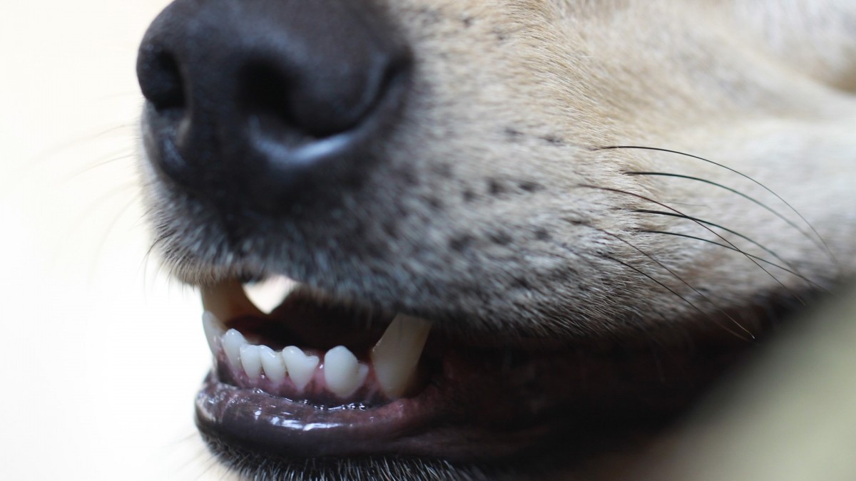
Analgesia and anesthesia are essential when obtaining dental radiographs. Ensure that the patient is adequately premedicated and then kept under a surgical plane of general anesthesia (i.e., unable to respond to stimuli) throughout the procedure. To avoid damage to the sensor, it should never be placed in the mouth of a patient who is not fully anesthetized and intubated. The tone of the jaw and the closing of the mouth can cause serious damage to the sensor. To avoid this damage, especially when it comes to dogs, it is recommended to place a bite block, a cut wide endotracheal tube, or a dental wedge between the molars on at least one side of the mouth.
4. Efficiency
Once the staff has achieved some training in this technique, speed will improve, but the technique should always be prioritized over speed, especially at the beginning.
5. Troubleshooting
It is important to have a thorough understanding of what can cause problems when obtaining dental X-rays, which can be boiled down to 3 variables:
A: animal.
B: beam.
C: sensor, phosphor plate, or film.
The technician must objectively measure each of the 3 parameters to obtain consistent diagnostic radiographs. When you get a non-diagnostic image, ask yourself what went wrong. The position of the animal's head (A), the angle of rotation of the beam (B), or the location of the sensor in the mouth (C)? Once the problem is identified, it must be corrected and tried again.
 Comments
Related Articles


Copyright © 2026 - All Rights Reserved
ISSN 2768-198X
List
Add
Please enter a comment