
Clinical Signs and Diagnosis of the Canine Primary Glaucomas

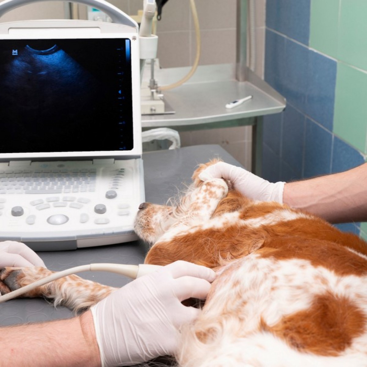
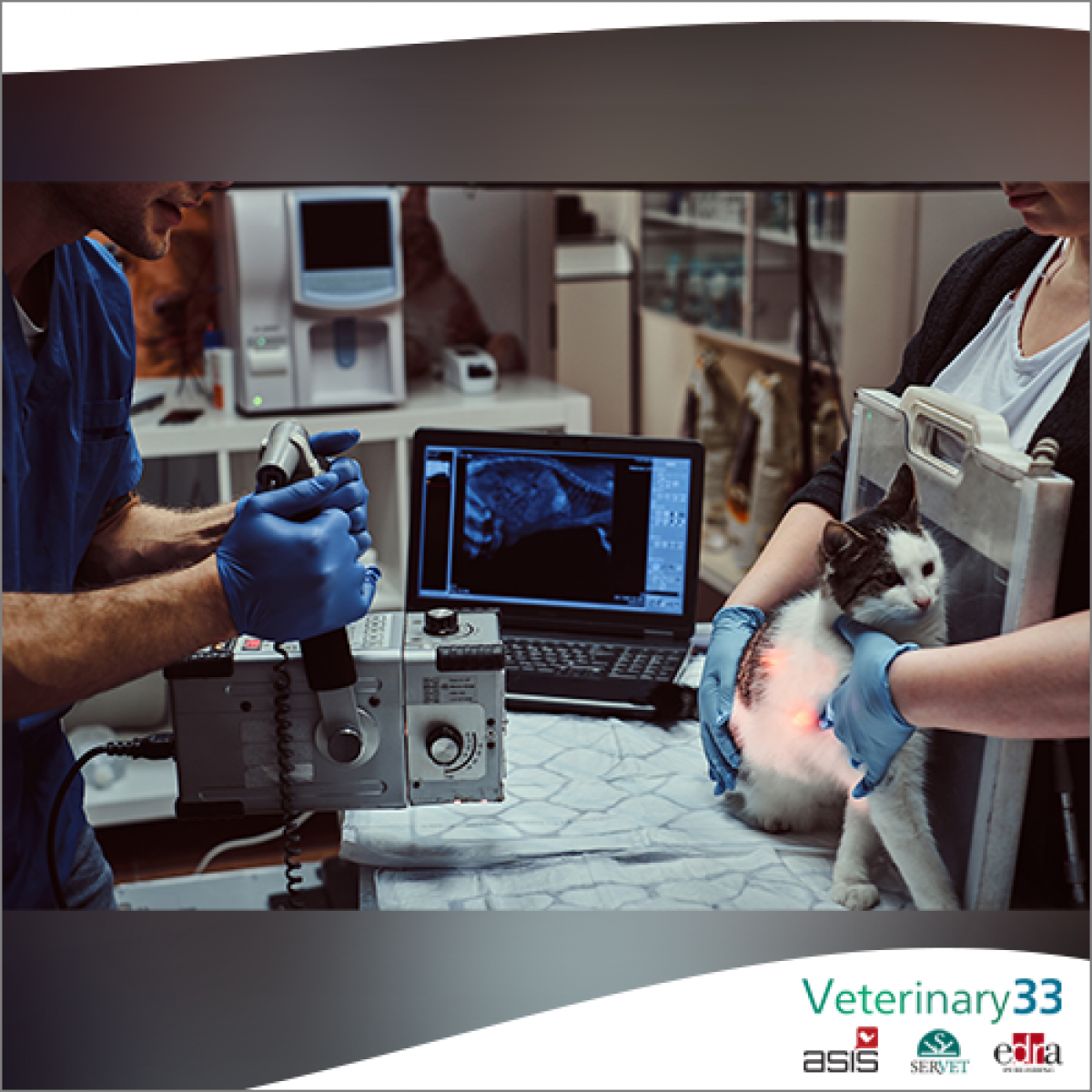
A clinician’s working definition of glaucoma determines which clinical signs and diagnostic values are considered important and pivotal in directing therapy. Unfortunately, there is no clear consensus on the definition of glaucoma or its various forms in human or veterinary medicine.1,2 For more than 100 years, glaucoma has been thought of as a “disease of elevated intraocular pressure (IOP)” and this parameter has been emphasized as the defining clinical sign, diagnostic variable, and therapeutic guide. This remains true for the vast majority of patients with glaucoma in veterinary medicine. Recent advances in both human and veterinary medicine, however, have demonstrated that damage to the optic nerve or associated tissues, such as the retinal nerve fiber layer (RNFL) can occur before IOP increases are documented or even after IOP has returned to normal values. This indicates that IOP alone is not the sole characteristic that defines the presence or absence of glaucoma and this fact has caused vision scientists to focus on the anatomic and molecular mechanisms that lead to the characteristic optic disc and visual field changes resulting in glaucomatous vision loss. The evolution of the definition of glaucoma to essentially a specific type of optic neuropathy has led to the proliferation of other diagnostic techniques that evaluate the posterior segment in more detail and tend to de-emphasize the role that IOP plays in this disorder.
For the purpose of this article, “primary glaucoma” is defined as a group of disorders that are typically bilateral, have a strong breed predisposition, are associated with increasing age, are documented or believed to have a genetic basis,3–5 result in characteristic changes to the optic nerve, and have no readily identifiable secondary ocular or systemic cause on routine ophthalmic examination. IOP is an important risk factor but not the sole risk factor for glaucomatous damage. The gonioscopic appearance of the iridocorneal angle (ICA) further categorizes primary glaucoma into “open” and “closed” forms. The term “primary angle closure glaucoma” (PACG) is used to reflect that the gonioscopic appearance of the ICA changes over time and eventually this structure appears closed.6,7 In clinical practice, PACG is many times more frequent than primary open angle glaucoma (POAG) in dogs and as such will be emphasized here.
Authors: Paul E Miller, Ellison Bentley
Source: https://pmc.ncbi.nlm.nih.gov/
 Comments
Related Articles

More news

Copyright © 2026 - All Rights Reserved
ISSN 2768-198X
List
Add
Please enter a comment