
Diagnosis and Surgical Correction of a Conjunctivopalpebral Dermoid in a Cat

Ocular malformations are rare anomalies of significant clinical relevance in veterinary ophthalmology. This case report aims to present the detailed diagnosis of a rare conjunctivopalpebral dermoid in a cat, supported by clinical and histopathological findings, and to describe the surgical intervention performed.
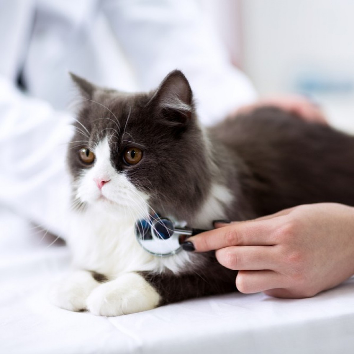
A 1.5-year-old male mixed-breed cat was referred to the Ophthalmology Clinic of Ankara University Faculty of Veterinary Medicine with complaints of a mass in the eye, pruritus, and discharge, which had been present since kittenhood. Clinical examination revealed a dermoid located on the right upper eyelid, affecting both the eyelid margin and the adjacent palpebral conjunctiva. Following the diagnosis of conjunctivopalpebral dermoid, the abnormal tissue was completely excised via surgical intervention. Histopathological examination confirmed the diagnosis, demonstrating keratinized epithelium, acanthosis, melanin pigmentation, hair follicles, sebaceous glands, and mast cells within the tissue.
No postoperative complications were observed, and the patient was monitored for approximately three months without any recurrence or additional issues. This report presents a comprehensive clinical and histopathological diagnostic approach and a successful surgical technique for a rare conjunctivopalpebral dermoid in a cat, offering a valuable contribution to the understanding and management of rare congenital anomalies in the field of veterinary ophthalmology.
Authors: Diğdem Uygur, Aslı Uygur, Arda Selin Tunç, Oytun Okan Şenel
Source: https://dergipark.org.tr/
 Comments
Related Articles


More news


Copyright © 2026 - All Rights Reserved
ISSN 2768-198X
List
Add
Please enter a comment