
Small ruminant dermatology review
by Jenny Alonge

Diagnosing and treating dermatologic disease in small ruminants can be challenging, because many dermatologic conditions, including certain foreign animal diseases (FADs), present similarly. Obtaining a detailed history, including farm details, previous disease outbreaks, and disease risk factors, is imperative to establish a diagnosis and to plan for effective management. The diagnostic investigation should also include collecting samples for cytology, culture and potentially histology. The following is a review of dermatologic conditions you may encounter when treating small ruminants.
Alpacas and llamas
Alpacas and llamas are commonly affected by ectoparasites, such as:
- Sarcoptes scabiei — Sarcoptic mange is particularly concerning, because the mites are a potential zoonotic agent. Lesions are typically crusted papules and pustules in the interdigital areas, medial thigh, ventral abdomen, thorax, axillae, perineum, and prepuce. Marked skin thickening and alopecia may occur in chronic infestations. Affected animals are typically extremely pruritic, and death has been reported in severe cases. Diagnosis is made by finding sarcoptic mites, eggs, or fecal pellets on a skin scraping, and by seeing mites on histologic examination. Treatment involves treating all herd animals for at least six weeks. Injectable avermectin products are recommended, because camelids tend to respond poorly to oral or topical treatments.
- Chorioptic sp. — Chorioptic mange has similar lesions and distribution as sarcoptic mange, but animals tend to be less pruritic. Chorioptic mites do not burrow and can survive for up to three weeks off the host. Topical treatments in conjunction with injectable medications are recommended, along with cleaning the living areas to reduce the mite burden.
- Zinc responsive dermatosis — Also known as idiopathic hyperkeratosis, this condition occurs in llamas and alpacas at any age. Lesions are non-pruritic papules with tightly adherent crusts found in the less densely haired areas, including the perineum, ventral abdomen, inguinal region, medial thighs, axilla, and medial forearms, and sometimes the face. Skin biopsy is necessary for diagnosis. Treatment involves administering zinc sulfate or zinc methionine and minimizing calcium supplementation.
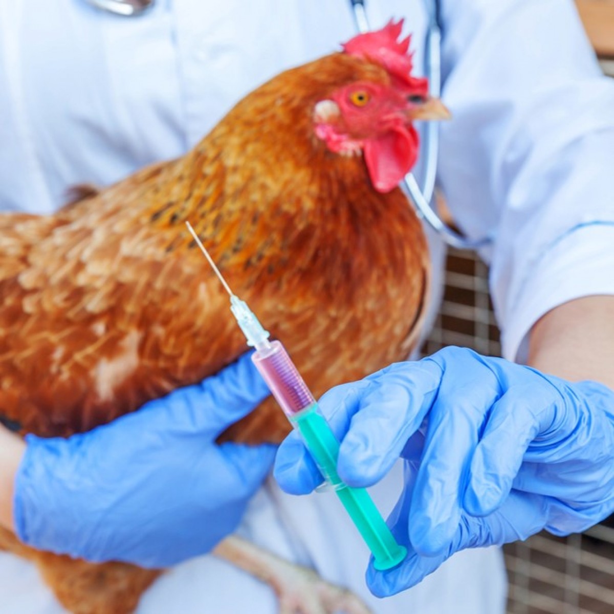
Sheep and goats
Numerous dermatologic conditions, including FADs, affect sheep and goats, including:
- Chorioptic sp. — Chorioptic mites are usually found on the distal limbs, especially around the pasterns. Topical treatments can complement systemic avermectin. In goats, topical avermectin treatment has variable absorption, depending on the breed, metabolic status, and subcutaneous fat. Double dosing is often required for effective treatment.
- Pemphigus foliaceous (PF) — PF in goats is one of the few autoimmune skin conditions recognized in livestock. The main clinical signs are generalized, severe pustular eruptions involving the entire body, crusting, and multifocal alopecia. Pruritus can also be significant. Outbreaks tend to be transient. Cytology can typically support diagnosis. Romanowsky staining of pustular material reveals acantholytic keratinocytes mixed with numerous neutrophils and in some cases, eosinophils. PF is a sterile pustular lesion and no bacteria should be seen in the polymorphonuclear cells. Secondary bacterial overgrowth can confuse diagnosis, so look for bacteria engulfed inside the cells before prescribing antibiotics. Definitive diagnosis requires histopathology. The PF treatment mainstay is injectable glucocorticoids, and long-term intervention is typically required. Topical glucocorticoid treatments are available, but do not seem effective against PF in goats.
- Culicoides hypersensitivity — Skin lesions and pruritus are present from spring to summer when Culicoides midges are most active. Seasonal recurrence is a key historical feature. The nonwooled body areas are affected, with lesions appearing as small crusts to marked skin thickening with ulceration and crust formation. Pruritus often manifests as foot stamping and suddenly dropping to the ground. Diagnosis is based on history and clinical presentation. Intradermal tests for Culicoides antigens are not formally validated diagnostic tests and the antigens are not standardized for any species. Management involves housing affected animals to reduce exposure when the insects are most active (i.e., from before dusk to after dawn). Applying topical insecticides and repellents may also be useful.
- Contagious ecthyma — Sheep and goats infected by the Parapox virus have lesions on their head, mouth, udder, genitalia, and limbs, and the esophagus and forestomach may also be affected. Some animals are subclinical carriers, and transmission may be facilitated by insects. Lesions can be painful, and kids and lambs may become lame or refuse to nurse, and dams who have lesions on their teats may not allow their young to nurse. Diagnosis is usually based on history and clinical signs. Treatment involves supportive care and pain control, but mild lesions typically resolve without treatment. While contagious ecthyma is highly contagious, mortality is usually low. The condition is zoonotic.
- Bluetongue — Bluetongue is a viral disease, family Reoviridae, genus Orbivirus, which has 27 serotypes worldwide. Culicoides midges are primarily responsible for transmitting bluetongue, most commonly in the late summer and early fall. Clinical signs vary from subclinical to rapidly fatal, and European fine-wool breeds tend to be more commonly and severely affected. Initial signs typically include fever, lameness, and reluctance to move, followed by hyperemic or ulcerated lips, muzzle, coronets, feet, and genitalia, generalized vasculitis, pulmonary edema, and facial and ear edema as the disease progresses. The tongue may become cyanotic, but this is uncommon. Abortions and birth defects may also occur in pregnant animals. Diagnosis involves testing via ELISA, quantitative pan or serotype-specific PCR, or virus isolation. Treatment is aimed at supportive care, including fluid therapy, nutritional support, antibiotics to treat secondary infections, anti-inflammatory medications, and pain control. Morbidity can be up to 100%, with 30% to 70% mortality rates. Bluetongue is a reportable disease.
- Dermatophilosis — Also known as rain scald or lumpy wool, this condition is caused by Dermatophilus congolensis, a gram-positive, aerobic actinomycete bacteria. Clinical signs include matted wool or hair in thick crusts that form paintbrush lesions, or raised lesions that resemble warts. Lesions can appear on any body area, but most commonly include the dorsum, distal limbs, ear tips, muzzle, pinnae, tail, udder, and scrotum. Elevating a crust and Gram staining a direct impression smear is usually diagnostic, revealing Gram-positive cocci in two to eight parallel “railroad tracks.” Treatment involves moving the animal to a dry environment, removing the crusts, applying topical treatment, and administering parenteral antimicrobial therapy. Dermatophilosis is zoonotic.
When addressing dermatologic disease in small ruminants, establishing a definitive diagnosis is critical to determine if the disease is reportable, and to devise an effective management strategy.
 Comments
Related Articles


More news


Copyright © 2026 - All Rights Reserved
ISSN 2768-198X
List
Add
Please enter a comment